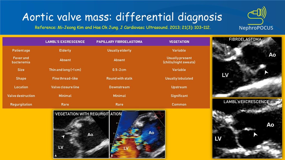
A complete workup showed no evidence of systemic infection but did reveal the presence of antiphospholipid antibodies. A papillary fibroelastoma is generally considered pathologically benign however outflow obstruction or embolism can be associated with syncope chest pain heart attack stroke and sudden cardiac death.
 Aortic Valve Papillary Fibroelastoma Associated With Acute Cerebral Infarction A Case Report
The radiology report thinks a healed vegetation is likely based on history of endocarditis I do not know where that comes from as I have never been diagnosed with this.

Aortic valve mass. When present on the aortic valve they are commonly confined to the ventricular aspect of the valve. A transesophageal echocardiogram showed a large highly mobile mass attached to the patients aortic valve. Symptoms due to papillary fibroelastomas are generally due to either mechanical effects of the tumor or due to embolization of a portion of the tumor to.
A 29-year-old asymptomatic young woman with incidental transthoracic echocardiographic TTE discovery of an aortic valve mass is presented. Join Leading Researchers in the Field and Publish With Us. Risk factors for adults include previous valve surgeries or a heart transplant calcium deposits in the mitral valve or in the aortic valve congenital heart defects or a history of endocarditis.
Advertentie International Journal of Inflammation Invites Papers on the Cell Biology of Inflammation. The sound occurs in the presence of a dilated aorta or in the presence of a bicuspid or flexible stenotic AV. Reis VS1 Tsang DC1 Williams DB1 Carrillo RG1.
Papillary fibroelastomas are rare benign excrescences with a propensity to arise on the surface of cardiac valves. Consisting of fibrous tissue covered by an elastic membrane the tumor is covered by the. If the cause of IE is injection of illicit drugs or prolonged use of IV drugs the tricuspid valve is most often affected.
Join Leading Researchers in the Field and Publish With Us. We discuss the differential diagnosis of a cardiac mass that includes infection tumor and thrombus. There was no improvement in the predictive model after indexing for aortic valve size or body size.
Aortic valve and mitral valve lesions may be somewhat more common than tricuspid valve and pulmonic valve lesions. The 2-dimensional TTE showed a mobile pedunculated mass attached by a thin stalk to the aortic surface of the right coronary aortic cusp at the junction of its base with the anterior aortic wall. Therefore additional procedures to accommodate a larger valve may be warranted in the aortic annulus smaller than 21 mm.
The lesions are pedunculated and consist of frondlike projections of connective tissue covered by endothelium. Aortic Valve Surgery Rankings Ratings US News Best Hospitals Rankings. The ejection click is a high-pitched sound best heard at the apex that occurs at the moment of maximal opening of the aortic valve AV shortly after the S1.
Subsequent TEE revealed a mass on the center cusp of my aortic valve. A complete resection of the tumor was achieved which was confirmed by postoperative TEE Figure 6. And aortic valve calcium mass scores.
Illicit drug use and IE. In this unique program specialists in valvular heart disease cardiac imaging interventional cardiology cardiac surgery and cardiac anesthesia work together to. The aortic valve was tri-leaflet and structurally and functionally found to be normal.
Massachusetts General Hospital in Boston MA. Advertentie International Journal of Inflammation Invites Papers on the Cell Biology of Inflammation. The aortic valve was found to be competent and functionally well without any residual defect or perforation after the procedure.
The mass is mobile. Subsequent coronary CT seems to rule out malignancy. The Heart Valve Program at the Massachusetts General Hospital Corrigan Minehan Heart Center provides a multidisciplinary team of experts to manage complex and common valve diseases.
Symptomatic Aortic Valve Mass - Cardiac Work-Up Challenges and Role of Computed Tomography Angiography. A score of 1850 AU captured 96 of patients with severe aortic stenosis iAVA. It disappears in patients with a severe calcific immobile aortic valve.
1University of Miami Miller School of Medicine Division of Cardiothoracic Surgery Miami Florida Division of Cardiothoracic Surgery University of Miami. In conclusion the current data suggest that the 19 mm SJM valve may not result in satisfactory left ventricular muscle mass regression despite adequate function even in small patients.